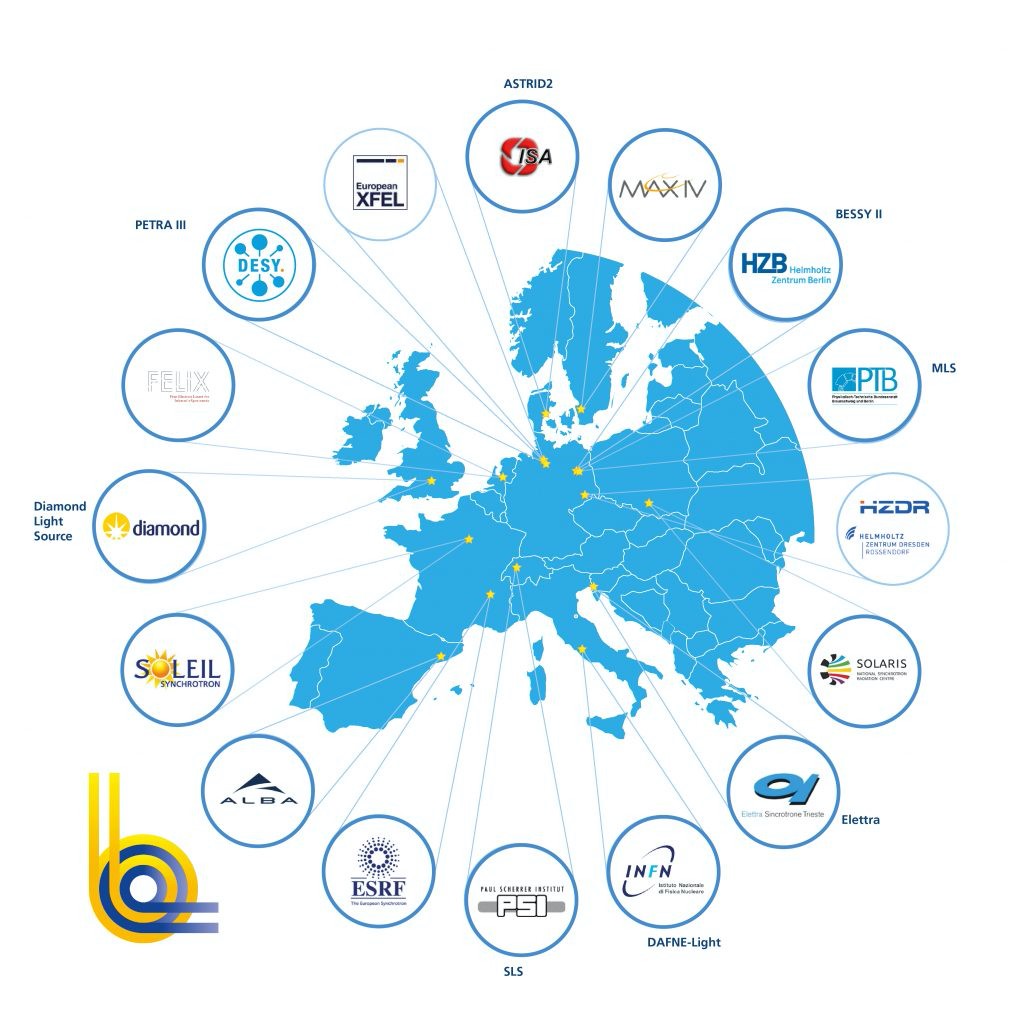
[Large Scale] - Introduction

Divider

[Synchrotron] - Introduction

Synchrotron radiation was first observed on April 24, 1947, at General Electric's research labs in the United States. At that time, synchrotron radiation was in fact viewed as an unwanted side effect, hampering the ambitions of physicists to reach ever higher energies in particle accelerators. Put simply, the charged particles in an accelerator emit radiation when travelling on a curved path at a velocity close to the speed of light. By emitting that so-called synchrotron radiation, the particles lose energy, hence their motion slows down. Synchrotron radiation is emitted within a narrow angle in the forward direction of motion of the charged particle. The synchrotron beam is thus highly collimated and and the desired wavelength can be selected from broad spectrum of the synchrotron radiation with a monochromator.

The usefulness of synchrotron radiation as an invaluable probe of matter was soon recognized. First experiments with synchrotron soft X-rays were conducted at Cornell University in 1956. Five years later, the first research program using synchrotron radiation was established at the National Bureau of Standards in the United States. Since then, synchrotrons improved at fast pace. Dedicated facilities have been built since the 1980s in which the focus resides on generating synchrotron light for research purposes. Medicine, materials science, biology, chemistry and geology have all greatly benefited from those developments.

Synchrotrons meant indeed a significant leap forward for research on the atomic structure of materials. But they have their limitations, too. Ultrafast processes crucial to many disciplines from biochemistry to materials science lie beyond their reach because light pulses produced at synchrotrons are not short enough. Due to their insufficient brightness, synchrotrons are also limited when it comes to the elucidation of the atomic structure of ultrasmall samples like nanostructures or some proteins.

From Waste Radiation to Light Sources
Over more than 60 years synchrotron radiation facilities have evolved from parasitic operation at particle accelerators to dedicated research tools. Since the first observation of synchrotron radiation, three generations of facilities have been developed.
A synchrotron is an extremely powerful source of X-rays. It builds on the physical phenomenon that a moving electron emits energy when it changes direction. If the electron is moving fast enough, the emitted energy, called synchrotron radiation, is at X-ray wavelength. Synchrotron radiation is emitted within a narrow angle in the forward direction of motion of the charged particle. The synchrotron beam is thus highly collimated and and the desired wavelength can be selected from broad spectrum of the synchrotron radiation with a monochromator.

Synchrotron radiation is millions of times more brilliant than any X-ray lab source. The technology provides opportunities to make new discoveries and products in fields such as materials, medicine and the environment.

Over more than 60 years synchrotron radiation facilities have evolved from parasitic operation at particle accelerators to dedicated research tools. Since the first observation of synchrotron radiation, three generations of facilities have been developed.
Various techniques are used in the experiments: imaging, spectroscopy and scattering, often combined together.

  • Imaging provides knowledge on what materials look like from the outside, as in a photograph, or from within, as in an X-ray image. The method delivers an image or film in two or more dimensions. The techniques used include microscopy and tomography. With nanometre resolution, it is possible to see an electronic component that has been built using nanotechnology. The technique can be used to see how the nanostructures within the component are affected when it is used, providing insights into how better and more efficient components can be built, such as more reliable catalysts.
  • Spectroscopy provides knowledge of chemistry and where the components in matter are positioned in relation to each other. Spectroscopy involves methods based on measuring the response that arises in the material when it is illuminated with various types of light. The techniques used include photoelectron spectroscopy and fluorescence. These techniques can be utilised to identify the chemical composition of an examined sample.
  • Scattering provides knowledge on the structure of atoms or molecules. Scattering is a term that covers various phenomena that arise when light meets a material. The light can be reflected or change direction when it passes through the material. These changes can be measured. The techniques used for scattering include X-ray crystallography and powder diffraction. These techniques can be used to identify how the atoms or molecules are positioned in relation to each other, which is important for the characteristics – mechanical, magnetic, electronic, etc. – of the matter or material. Scattering can be used to observe battery materials in order to see how the atoms move when the battery is charged and thereby gain a better understanding of how improved, lighter and cheaper batteries can be designed. It is also possible to see how the structure of a material changes when it is subjected to mechanical forces. This provides knowledge that can be used to develop new, stronger materials with higher breaking strength.

Experiments at Large Scale Facilities are performed upon research proposal acceptance. Deadlines for proposal submission are normally 2-3 times per year, depending on the Facilities. Proposals are peer reviewed by expert panels, graded and accepted for beamtime allowance only if they meet cut-off criteria. Before writing a proposal, you must clearly identify the instrument(s) and the instrument requirements for your needs. Contact the instrument responsibles and discuss your experimen feasibility and all related technical issues.

Writing a proposal always specify:

  • Proposal Summary
  • Aims of the experiments and background (scientific background)
  • Experimental Methods (measurement strategy)
  • Beamlines and Beamtime requested
  • Results Expected
  • References

[Large Scale] - Top page grid

Divider

[X-FELS] - Introduction

Synchrotrons meant indeed a significant leap forward for research on the atomic structure of materials. But they have their limitations, too. Ultrafast processes crucial to many disciplines from biochemistry to materials science lie beyond their reach because light pulses produced at synchrotrons are not short enough. Due to their insufficient brightness, synchrotrons are also limited when it comes to the elucidation of the atomic structure of ultrasmall samples like nanostructures or some proteins.That is why scientists want to take the next step in light source technology.

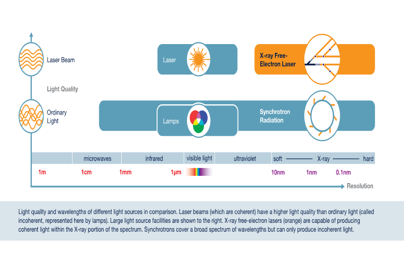
Challenges like capturing the single steps of a chemical reaction by seeing how atoms move and bonds are broken up, while new ones are forged calls for a more powerful and faster new generation of X-ray sources. This new generation is represented by so-called X-ray Free Electron Lasers (FEL), the 4th generation of X-ray light sources. X-FELs feature a brilliance, 1 billion higher than that of the most advanced synchrotrons and they can produce ultrashort pulses of coherent and polarized light.

development of X-FELS

X-FELS pulses are:

  • extremely bright (109 times brighter than synchrotron X-rays)
  • extremely short (femtoseconds)
  • coherent

Thanks to these features listed above, X-FELs will be able to reveal the structure of membrane proteins which are the key to more rational drugs against diabetes and heart disease.
Ultrafast processes of interest in chemistry will become accessible to the ability of FELs to produce very short pulses of X-rays. In addition, magnetic materials for new information storage technologies could be explored with the polarized beams of FEL light. Many other scientific and technological benefits are imaginable in other areas and the full potential of X-FELs will only be unveiled when scientists start using them. It is already clear at this point in time that they will open new avenues of research while complementing already existing techniques such as those applied at synchrotrons, neutron or muon imaging facilities.

features of X-FELS


Experiments at Large Scale Facilities are performed upon research proposal acceptance. Deadlines for proposal submission are normally 2-3 times per year, depending on the Facilities. Proposals are peer reviewed by expert panels, graded and accepted for beamtime allowance only if they meet cut-off criteria. Before writing a proposal, you must clearly identify the instrument(s) and the instrument requirements for your needs. Contact the instrument responsibles and discuss your experiment feasibility and all related technical issues.

Writing a proposal always specify:

  • Proposal Summary
  • Aims of the experiments and background (scientific background)
  • Experimental Methods (measurement strategy)
  • Beamlines and Beamtime requested
  • Results Expected
  • References

[Large Scale] - Top page grid

Divider

[Neutron] - Introduction

To determine the positions and motions of atoms in condensed matter

Neutron Advantages

  • Wavelength comparable with interatomic spacings
  • Kinetic energy comparable with that of atoms in a solid
  • Penetrating ⇒ bulk properties are measured & sample can be contained
  • Weak interaction with matter aids interpretation of scattering data
  • Isotopic sensitivity allows contrast variation
  • Neutron magnetic moment couples to B ⇒ neutron “sees” unpaired electron spins

Neutron Disadvantages

  • Neutron sources are weak => low signals, need for large samples etc
  • Some elements (e.g. Cd, B, Gd) absorb strongly
  • Kinematic restrictions (can’t access all energy & momentum transfers)

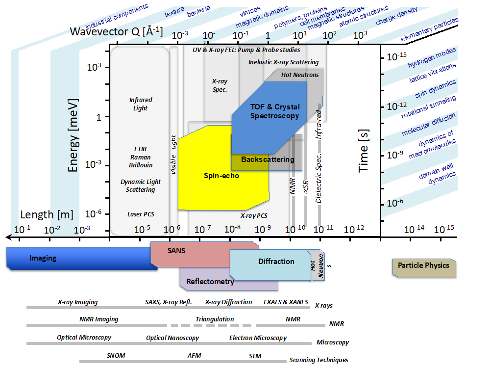
The figure below shows the time and length (energy and wavevector) scales of the various neutron-based techniques (Courtesy of Ken Andersen - ESS). You can use it to identify the techniques which will best match your needs and then consult the list below to choose the appropriate instruments.

  • Neutron diffraction reveals structural information on the arrangement of atoms and magnetic moments in condensed matter.
  • Small Angle Neutron Scattering (SANS) explores the mesostructures of liquids and solids on length scales ranging from 1 nanometre to about a micron.
  • Neutron reflectometry gives information (depth-dependent composition) on the structure of thin films and surfaces which can be solid/solid, solid/liquid, liquid/liquid and liquid/air interfaces.
  • Neutron spectroscopy (TOF, TAS, Spin-echo, Backscattering) probes the dynamics of magnetic moments, molecules and lattices over length scales ranging from a few angstroms to tens of nanometers, and over timescales from tens of picoseconds up to the microsecond.
  • Neutron imaging  is a non-destructive technique, highly complementary to X-ray imaging, that can see inside materials and examine processes therein.

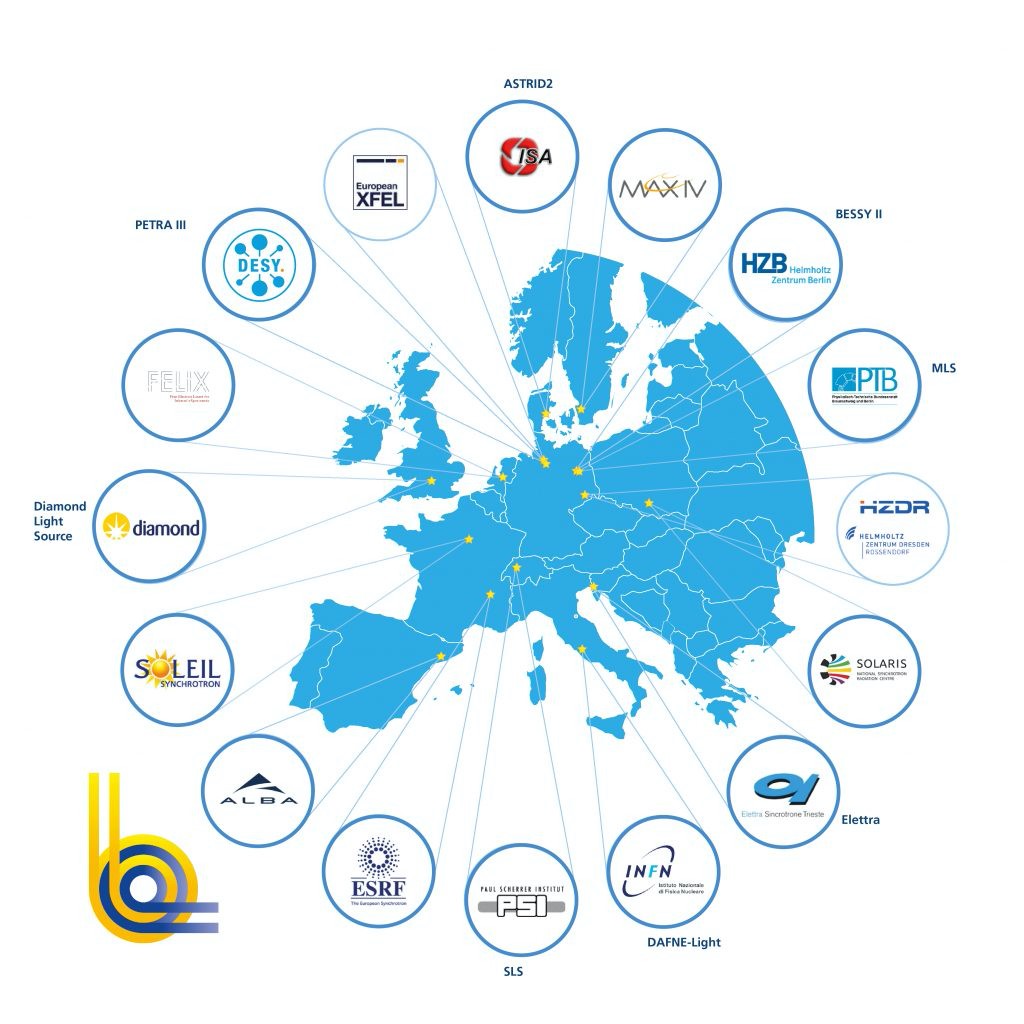
Neutrons map

Experiments at Large Scale Facilities are performed upon research proposal acceptance. Deadlines for proposal submission are normally 2-3 times per year, depending on the Facilities. Proposals are peer reviewed by expert panels, graded and accepted for beamtime allowance only if they meet cut-off criteria. Before writing a proposal, you must clearly identify the instrument(s) and the instrument requirements for your needs. Contact the instrument responsibles and discuss your experimen feasibility and all related technical issues.

Writing a proposal always specify:

  • Proposal Summary
  • Aims of the experiments and background (scientific background)
  • Experimental Methods (measurement strategy)
  • Beamlines and Beamtime requested
  • Results Expected
  • References

[Large Scale] - Top page grid

Divider

[Large Scale] - Learn more section text

[Large Scale] - Menu aside